More about active mobilzation

Given normal, healthy joint surfaces, full range of movement depends on the extensibility and mobility of the soft tissue structures. This is particularly true of movements that require elongation of myo-fascial-neural (M-F-N) structures over more than one joint. 

Short duration (minutes)  passive  stretching the M-F-N structures does not usually give an immediate increase in the range of movement. However, active stretching techniques such as the PNF hold relax technique,  have  been shown to produce an immediate increase in range of movement.  The principle of the hold-relax technique is to induce an isometric  contraction of the targeted muscles in the lengthened range . 
 
Active mobilisation uses the same basic principle – lengthened muscles are made to contract isometrically.  The difference is that the isometric contraction is not maximal but rather consists of several repetitions of  sub-maximal isometric contractions of the target and the surrounding muscles. 
 
The underlying mechanism promoting an increase in range of movement probably relates to changes in the visco-elastic behaviour of the areolar tissue that lies between layers of fascia in the myo-fascial-neural system. 

The basic technique

  1. In active mobilisation the limb segments are passively moved to a position where the target muscles and related fascial structures are lengthened (stretched).
  2. The limb position is then maintained by an external force (manual holding by the therapist) during several cycles of contraction – relaxation of  the target and surrounding muscles.
  3. If the techniques is effective there will be an increase in the range of movement following the application of the active stretch.
  4. Stretching is followed by activities that involve the tight muscles in their lengthened range of movement.

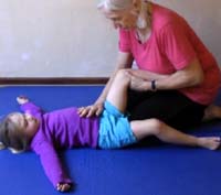
Active mobilization to increase straight leg raise (SLR)

Structures that limit SLR include: 

The structures limiting straight leg raise include:  lumbodorsal fascial, gluteal fascia, iliotibial band, fascia lata, hamstring muscles and their related  fascial extensions, neurovascular bundle related to the sciatic nerve. 

Hip elongation in flexion
The child lies in supine. The right hip and knee are flexed to a position that places a mild stretch on the posterolateral fascial system. 

roan hip in supine.jpg

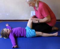
The therapist holds the limb in this position and applies a longitudinal force along the long axis of the femur which stretches the shorter structures around the hip joint. 

The therapist maintains the limb in this position for the duration of the cycle of contraction-relaxation of the target muscles.  

roan hip in supine 1.jpg

The child is instructed to lift and lower the left lower limb keeping the knee straight  to 50-6010 times at a pace of about  about 4s per up and down movement of the limb. This movement activates the hip extensors on the left and the child actively pushes down against the therapist's hand. 
 

roan hip in supine 2.jpg


Video demonstration